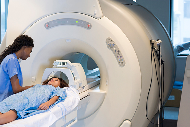
This article will delve into the various aspects of radiotherapy in pediatric cancer, a crucial treatment approach for children battling cancer. We’ll explore how this specialized therapy works, the different methods of delivery, the dedicated team involved in a child’s care, and what families can expect throughout the treatment process, including potential side effects and their management.
What is Radiotherapy?
Radiotherapy is a medical treatment that utilizes high-energy radiation, such as X-rays, to target and destroy cancer cells within the body. This approach aims to either shrink or eliminate tumors. It can be employed at various stages of a child’s cancer treatment, with the specific timing and number of sessions varying based on the treatment’s objective, the type of cancer, and the child’s age. When a child is referred for this therapy, the medical team provides detailed information about their personalized treatment plan.
The fundamental principle behind radiotherapy is that the radiation damages the DNA inside cancer cells, causing them to die or lose their ability to multiply. While healthy cells also experience damage from radiation, they possess a greater capacity to recover from it compared to cancer cells. This is why radiotherapy is typically administered in multiple smaller doses over time. This approach allows healthy tissues to mend between sessions, while cancer cells accumulate irreparable damage and are eventually destroyed.
However, it’s important to understand that exceeding certain radiation doses can lead to permanent harm to healthy tissues. Each type of cancer, along with various healthy organs and tissues, responds differently to radiation. Consequently, a child’s radiotherapy plan is meticulously designed to deliver the optimal radiation dose directly to the cancer cells while significantly reducing exposure to the surrounding healthy areas.
Radiotherapy in Pediatric Cancer
Children may undergo radiotherapy for several key purposes. One reason is to reduce the size of tumors before surgical removal, a practice known as neoadjuvant radiotherapy. Another common application is to eliminate any remaining cancer cells near the original tumor site following surgery, thereby decreasing the likelihood of recurrence; this is referred to as adjuvant radiotherapy. In situations where a cure is not achievable, radiotherapy can be used to alleviate symptoms and enhance the child’s comfort, forming a component of palliative care.
Additionally, radiotherapy may be used to prepare a child’s body for a stem cell transplant, a procedure for certain blood cancers. In this scenario, it is delivered to the entire body, known as total body irradiation, primarily to suppress the immune system and prevent the rejection of donor blood stem cells after transplantation.
How Radiotherapy Functions
Regardless of the specific type, all radiotherapy modalities operate by damaging the DNA within cancer cells, which ultimately causes them to die or prevents them from multiplying. A critical distinction lies in how healthy cells and cancer cells respond to this damage. Healthy cells possess a remarkable capacity to recover significantly, or even completely, from a limited amount of radiation exposure, whereas cancer cells are intended to suffer permanent damage and be eradicated. For this reason, radiotherapy is typically divided into a series of treatments.
This allows healthy cells time to recuperate between sessions, while the cancer cells progressively accumulate damage, leading to their demise. However, it is important to recognize that exceeding certain radiation doses can result in permanent harm to healthy tissues. The sensitivity to radiotherapy varies among different cancer types, as well as among healthy organs and tissues. Consequently, each child’s radiotherapy plan is meticulously crafted to ensure the optimal delivery of radiation to the cancer cells while safeguarding as much of the surrounding healthy tissue as possible.
External Beam Radiotherapy
This is the most common form of radiation delivery, where a machine directs radiation beams from outside the body towards the tumor.
- Photons (High-Energy X-rays): Most radiotherapy centers primarily use photons, which are high-energy X-rays generated by a linear accelerator. The invisible photon beam travels through the body, delivering a dose to the tumor and the tissues it traverses. As the linear accelerator rotates around the patient, it shapes the photon beam, ensuring that the highest radiation dose is concentrated precisely at the tumor’s location and conforms to its shape, while surrounding tissues receive only lower doses.
- Protons (Charged Particles): Proton therapy centers utilize high-energy protons as external radiotherapy. These charged particles are accelerated to very high speeds for beam delivery, which is similar to photons in that a machine can rotate around the patient, and the beam is invisible and unfelt. The primary advantage of protons is their controlled penetration; the beam delivers its dose within a defined area and then stops, delivering very little or no radiation beyond that point. This characteristic allows for a significant reduction in radiation dose to healthy normal tissues compared to photons. Children are carefully selected for proton beam therapy based on the tumor’s size and position in the body, and whether this approach is expected to result in fewer long-term side effects compared to photon treatment.
Internal Radiotherapy
Less commonly used, internal radiotherapy involves administering radioactive materials directly within the body.
- Brachytherapy: This is a localized form of internal radiation. It involves inserting several very thin, flexible plastic tubes directly into or around the tumor or its cavity, sometimes during an operation to remove the tumor. These tubes remain in place for the duration of the treatment course, which can range from 3 to 7 days. After placement, a scan is performed to create an individualized radiation treatment plan. The brachytherapy dose is delivered by connecting the implanted tubes to a machine that guides a tiny radioactive source into precisely planned locations within the tubes. Once complete, the source retracts, and the tubes are disconnected. Treatment is given multiple times a day over consecutive days, necessitating a hospital stay. The positioning of the tubes is meticulously checked before each session to ensure accuracy, and after the final treatment, the medical team removes the tubes.
- Molecular Radiotherapy: This involves administering radioactive drugs either orally or intravenously. The substance then circulates through the bloodstream and is selectively absorbed by tumor cells, regardless of their location in the body. While there are only a limited number of indications for molecular radiotherapy in children, it can be a valuable or even curative treatment, even for widespread disease. An inpatient stay in a specially protected room is required, and parents and staff must adhere to safety guidelines to minimize their personal radiation exposure.
Precision in External Radiotherapy Delivery
When a child undergoes external radiotherapy, utmost precision is paramount. This involves meticulously defining the target area, reliably identifying any nearby healthy tissues, accurately directing the radiation dose to the target region, and minimizing the dose to adjacent organs at risk of long-term side effects. This intricate process necessitates planning scans performed shortly before radiotherapy begins, often reviewed in conjunction with other scans from earlier in the illness. Radiotherapy planning is a careful and time-consuming process, typically spanning several days to two weeks. Even in palliative settings where treatment may begin on the same day, several hours are needed to create a plan.
The radiotherapy plan is developed using a planning CT scan, acquired with the child in the exact position they will maintain during treatment. Therefore, it is crucial for the child to remain very still during radiation delivery, replicating the planning scan position. Immobilization devices may be required, and anesthetic sedation is frequently necessary for very young children. To ensure consistent patient positioning, several checks are performed before each radiotherapy session. Modern planning and verification technologies are employed to ensure the treatment is as precise and safe as possible.
The Radiotherapy Team and Their Roles
A comprehensive team of healthcare professionals collaborates closely to provide optimal care for children receiving radiotherapy.
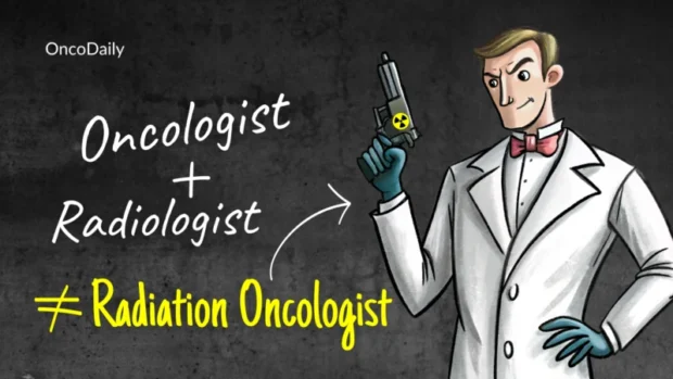
Radiation Oncologist
This physician is responsible for planning and overseeing the radiation treatment. They are an integral part of the pediatric oncological medical team that collectively discusses and monitors the child’s entire treatment program. During initial visits to the radiotherapy department, the radiation oncologist explains the treatment, its potential benefits, and anticipated side effects, and obtains the family’s consent. After the planning scan, they work with the radiotherapy team to construct the treatment plan. Throughout the treatment period, the child has regular consultations with the radiation oncologist to monitor and manage side effects. The radiation oncologist may also be involved in follow-up visits post-treatment.
Radiotherapy Technologists (RTTs) / Therapeutic Radiographers
These registered healthcare professionals manage the patient’s daily radiotherapy treatment pathway under the radiation oncologist’s supervision. Their duties include ensuring accurate patient positioning for treatment, selecting appropriate immobilization equipment, acquiring the necessary radiotherapy planning scans, and utilizing their technical expertise for precise treatment delivery.
Health Play Specialist
A crucial member of the team, Health Play Specialists are instrumental in preparing a child for radiotherapy. They employ play techniques, videos, and demonstrations to explain what the child can expect throughout their treatment journey. This is especially vital in helping children process and cope with any anxieties or feelings they may have regarding the treatment.
Dosimetrist
Once the radiotherapy planning scan is completed and the radiation oncologist has defined the area to be treated, the dosimetrist begins to create an individualized radiotherapy treatment plan. Dosimetrists work directly with the radiation oncologist to design the optimal radiation dose for treatment.
Clinical Physicist
The clinical physicist meticulously verifies the dosimetrist’s work, employing numerous checks and software programs to assess the safety of each treatment plan. They are also responsible for ensuring that all medical equipment and software are used safely. Although patients rarely interact with the dosimetrist and clinical physicist directly, these professionals are pivotal in the development of a radiotherapy treatment plan.
Anesthesiology Team
In situations where a child, especially a very young one, may not be able to remain still for the duration of a radiotherapy appointment, patient sedation or general anesthesia may be considered. The anesthesiology team manages the sedation process, collaborating closely with the therapeutic radiographers to carry out the necessary procedures.
Other Health Professionals
Depending on the structure of the radiation oncology department or pediatric oncology clinic, a child may have appointments with other healthcare professionals. These specialists, such as nutritionists, physical therapists, psychologists, dentists, and social workers, address specific needs and provide guidance to the child and family throughout the treatment process.
The Radiotherapy Process: From Consultation to Follow-up
The journey of pediatric radiotherapy involves several distinct stages, each designed to ensure precise and safe treatment.
Initial Consultation
During the first visits to the radiotherapy department, the family will meet the radiation oncologist, who will explain the treatment plan and its preparation. This discussion will cover potential side effects that may occur during and shortly after treatment, as well as those that might appear years later. The verbal explanation may be supplemented with written information. The doctor may examine the child and ask questions, and will seek informed consent for treatment once the family fully understands the proposed plan. It can be beneficial to write down questions before this appointment. The doctor may also discuss whether general anesthesia is recommended, particularly for young children or patients who may struggle to remain still.
Mould Room and Simulation
To ensure treatment accuracy, it is absolutely essential for the child to remain still during the radiotherapy planning scan and subsequent treatment sessions. To aid in maintaining stability and reproducible patient positioning, an appointment in the mould room may be required before the planning scan. Here, radiographers assess the child’s positioning and determine if any immobilization equipment is needed for treatment. While immobilization equipment helps reproduce patient positioning and enhance treatment accuracy, the specific choice varies based on the treatment site and departmental protocols.
The treatment planning phase begins with simulation, which involves acquiring a scan to precisely show the target region while the child is in the treatment position. CT, PET-CT, and MRI scans may be used for planning purposes, with the radiation oncologist determining the most suitable scan for each individual case. Radiographers may take time to reassess the child’s positioning before the scan and may use pens to draw temporary marks on the skin. After the scan, they might use a needle and ink to create small, freckle-like permanent dots in the previously marked areas.
Depending on the treatment site and departmental procedures, a child may have two or three of these dots, which assist in accurate and reproducible positioning for treatment. The radiation oncologist may also request the administration of intravenous contrast for the planning scan. Occasionally, the planning scan is repeated during treatment, for example, if there are changes due to reduced post-operative swelling or other anatomical shifts.

source: www.oncologynurseadvisor.com
Treatment Planning
Following the simulation, the radiation oncologist collaborates with dosimetrists and radiation physicists to develop the individualized treatment plan. This team determines the precise amount and frequency of radiation and meticulously maps the radiation beams. The radiation oncologist issues a prescription outlining the exact radiation dose, frequency, and location. The treatment plan is unique to each child, depending on various factors such as the tumor type and the child’s age. Before the first treatment, the radiation therapy team takes X-ray images on the treatment machine to confirm the patient’s positioning and verifies that all machine settings are correct. These images ensure the treated area is precisely as planned by the doctor, and the images must be approved before treatment commences.
The radiation oncologist will discuss the finalized treatment plan with the family before treatment sessions begin, providing an excellent opportunity for questions. A child life specialist may also work with patients to explain the treatment process using child-friendly language and medical play.
The radiation oncologist will outline the approximate duration of each session, the total number of treatments (often five days a week for one to seven weeks), and the number of “rest” days between treatments, which allow cancer cells to die and healthy cells to recover. They will also emphasize the importance of the child remaining still during treatment, and child life specialists can help create a coping plan. If a child cannot stay still, general anesthesia may be employed, involving an anesthesiologist as part of the treatment team.
Typical Radiotherapy Appointment
Most patients receive radiation treatment on an outpatient basis. The scheduler coordinates appointments with families, noting that general anesthesia procedures will have a separate appointment. Before each visit, patients and parents check in and proceed to the radiation oncology waiting area, which often includes a play area in pediatric centers. When it is time for treatment, a member of the care team, usually a radiation therapist or nurse, escorts the patient and sometimes a parent to the treatment area. If general anesthesia is required, a member of the anesthesia team will meet the patient and parent. At some centers, a parent can remain with the child until treatment begins, while others may ask parents to wait outside.
In the treatment room, the radiation therapist or nurse positions the patient on the treatment table using their mask or body mold, ensuring comfort with items like blankets or cushions. This positioning phase can sometimes take longer than the actual radiation delivery. Once the patient is positioned, the radiation therapist moves to an adjacent or nearby room to initiate the treatment. The child remains alone in the treatment room, but the therapist can see, hear, and communicate with them at all times.
The machine will align to precisely target the radiation, sometimes by taking an X-ray image for confirmation. Laser beams are used for positioning but are not felt. The child will not feel or see the radiation treatment being delivered, though the machine will make noise. After the session, the care team helps the patient off the bed. If anesthesia was not used, children can typically resume normal activities. If anesthesia was administered, the child must recover before leaving the radiation oncology area, with recovery time varying based on their individual response.
Follow-up Visits
Following the completion of therapy, the patient will have regular follow-up visits with the radiation oncologist. These visits often include diagnostic imaging tests, such as CT, PET, or MRI scans, to monitor the cancer’s response to treatment.
Helpful Tips for Families
To ensure smooth treatment, arriving on time or even a few minutes early for appointments is advised to allow for check-in and avoid delays for other patients. If a mask is used, the child’s hairstyle should remain the same as when the mask was made; a haircut before simulation may be necessary if hair loss is anticipated.
Loose, comfortable clothing without metal, such as T-shirts with sweatpants or elastic-waist pants, is recommended, or the patient may wear a hospital gown. Pediatric centers have varying policies regarding parental observation during therapy sessions; some allow one or both parents, while others require parents to wait. Siblings are generally not permitted to observe and must be supervised by an adult in the waiting area, so families should plan accordingly.
Radiotherapy Side Effects
Radiotherapy can lead to various physical changes because the radiation affects healthy cells alongside cancer cells. The occurrence and severity of side effects depend on several factors: the specific body area treated, the total duration of the radiation treatment, the daily radiation dose, the overall radiation dose, any pre-existing health issues, and other concurrent treatments. Predicting exact outcomes is difficult as individual responses vary. Some children experience significant side effects impacting daily life, while others have only minor complaints and can continue with routine activities. Your child’s radiation oncologist can provide more precise information about the individualized plan and expected side effects.
Short-Term Side Effects
These temporary side effects typically appear during treatment and for a few weeks afterward.
- General Complaints: Fatigue or low energy is a common general complaint that can persist for a while after treatment concludes. The degree of energy loss varies among children, and some may not be significantly bothered by it. There are no strict lifestyle rules; activities should be adjusted to the child’s energy level. Any prohibitions on activities like swimming or contact sports during or after therapy should be discussed with the doctor.
- Skin Reaction and Care: External radiation passes through the skin, which can react with redness, usually appearing two to three weeks after the first treatment. The skin may also feel itchy or tender. These reactions might worsen for 7 to 10 days after the last treatment but typically recover within 2 to 4 weeks. Hair loss from the irradiated skin can occur but usually regrows, depending on the dose. Special care for the irradiated skin is crucial during and after radiotherapy: showers are preferred over baths (also to preserve skin markings), swimming should be avoided if the skin is irritated, non-permanent markings should not be removed until after the last treatment, and exposure to extreme heat or cold should be avoided (e.g., heating pads, ice packs, hot tubs).
The skin should be washed with mild, pH-balanced, unscented soap (like baby shampoo) and patted dry gently, not rubbed or scrubbed. Shaving the treated area is not recommended, nor is using deodorants or antiperspirants if the armpits are irradiated. Tight or abrasive clothing, scratching, and applying band-aids, tape, perfumes, oils, lotions, salves, or unauthorized creams should be avoided. The irradiated skin is susceptible to sunburn and must be protected from the sun with clothing and sunscreen (SPF 30 or higher) even after healing. The radiotherapy team will advise on skin care and specific creams or bandages to use, and they should be consulted if signs of infection (e.g., increased swelling, redness, blistering, pain) appear.
- Hair Loss: If the irradiated area has hair growth, hair loss can occur, most noticeably on the scalp or eyebrows if the head is treated. Hair usually begins to regrow a few months after radiotherapy, though its texture or color might change. In some cases, hair loss can be permanent, depending on the radiation dose and other therapies affecting hair growth. The radiotherapy team can advise on scalp care and options for covering the head, such as wigs, hairpieces, headbands, caps, or hats.
- Eating and Drinking: A child’s appetite might decrease during radiation treatment. Generally, maintaining a healthy, balanced diet is advised to sustain energy levels. If a dietitian is involved, a dietary plan should be discussed, and any restrictions should be clarified with the radiation oncologist. If nausea or vomiting occurs, anti-nausea medication may be prescribed and should be kept at home. If the medicine is ineffective, the care team should be contacted.
- Reduction in Blood Cells: Depending on the treated area, radiotherapy can sometimes affect the bone marrow, where blood cells are produced. This could lead to increased bruising or bleeding, a higher risk of infections, or anemia.

Read OncoDaily’s Special Article About Radiation Oncologists
Long-term and Late Side Effects of Radiotherapy
These side effects may become apparent months or even years after a child’s radiotherapy treatment has concluded, and they often represent permanent changes.
Developmental and Cognitive Challenges
Radiotherapy applied to the brain can significantly influence a child’s physical and mental growth and development. This may result in cognitive difficulties, impacting thinking processes and potentially leading to learning disabilities.
Endocrine System Deficiencies
Children may experience central endocrine deficiencies, which involve the impaired production of essential hormones. These hormones are critical for various bodily functions, including growth, the onset of puberty, fertility, weight regulation, and the proper functioning of the adrenal and thyroid glands. Specifically, thyroid problems can also emerge if radiation is delivered to the head, neck, or chest areas.
Vascular and Sensory Issues
Problems affecting blood vessels can be a concern in diverse areas that have received treatment, such as the brain, head and neck, chest, abdomen, pelvis, and limbs. Furthermore, radiotherapy to the brain, head, or neck can lead to impairments in hearing or vision, and may contribute to eye problems like cataracts.
Bone and Growth Impact
Radiation therapy has the potential to hinder bone growth, particularly in developing areas like the face, skull, spine, ribs, pelvis, and the long bones of the arms and legs. This can result in spinal issues, such as kyphoscoliosis.
Organ-Specific Complications
Irradiation of certain body regions carries risks for specific organs. If the chest or abdomen is treated, children may experience lung and breathing difficulties, as well as heart problems. Abdominal or pelvic radiation can lead to kidney or liver issues, along with problems affecting the bowel or digestive system. Spleen dysfunction is a possible complication when the abdomen is irradiated.
Fertility and Reproductive Health
Treatment that involves the pelvic or groin area can impact the testicles and ovaries, even in young children, potentially reducing their fertility later in life. Concerns about future pregnancies are also relevant if the abdomen or pelvis receives radiation. For some children, discussions about egg or sperm banking may be recommended to preserve future reproductive options.
Neurological and Tissue Changes
Radiation to the pelvis can result in problems with nerve tissue. Additionally, the skin in treated areas, particularly the pelvis and extremities, may develop fibrosis, which is a hardening of the tissue.
Permanent Hair Loss
Depending on the radiation dose delivered, hair loss in treated areas such as the head, neck, or chest can become permanent.
Risk of Secondary Cancers
Although uncommon, a long-term consequence of radiation therapy can be the development of a new type of cancer within the previously irradiated area. This potential risk underscores the importance of ongoing regular follow-up visits after therapy is completed. Healthcare teams diligently monitor for signs of secondary cancers, such as changes in the skin, alterations in moles, bone pain, or the appearance of persistent thickening or lumps during physical examinations. Patients, or their parents if the child is very young, are also encouraged to perform monthly self-examinations.
Long-term and Late Side Effects of Radiotherapy
The enduring effects of radiotherapy on children, often appearing months or even years after treatment, can be permanent.
Developmental and Learning Impacts
When the brain is treated with radiation, it can significantly affect a child’s physical and mental development. This may lead to cognitive challenges, impacting their thought processes and potentially causing learning disabilities.
Hormone Production Issues
Children might experience central endocrine deficiencies, meaning their bodies produce insufficient amounts of crucial hormones. These hormones are vital for normal growth, pubertal development, fertility, weight management, and the proper function of their adrenal and thyroid glands. Specifically, radiation to the head, neck, or chest can also result in thyroid problems.
Blood Vessel and Sensory Complications
Issues with blood vessels can arise in various areas that have received radiation, including the brain, head and neck, chest, abdomen, pelvis, and limbs. Furthermore, treatment to the brain, head, or neck can impair hearing or vision, and may contribute to eye problems like cataracts.
Effects on Bones and Growth
Radiotherapy has the potential to hinder bone growth, particularly in developing bones found in the face, skull, spine, ribs, pelvis, and the long bones of the arms and legs. This can sometimes lead to spinal problems, such as a curvature known as kyphoscoliosis.
Organ-Specific Challenges
Radiation to certain body regions carries distinct risks for specific organs. If the chest or abdomen is treated, children might experience lung and breathing difficulties, along with heart problems. Radiation to the abdomen or pelvis can result in kidney or liver issues, as well as problems with the bowel or digestive system. Additionally, the spleen may become dysfunctional following abdominal radiation.
Fertility and Reproductive Health
Treatments involving the pelvic or groin area can affect the testicles and ovaries, even in very young children, potentially reducing their fertility later in life. Concerns about future pregnancies are also relevant if the abdomen or pelvis receives radiation. In some cases, discussions about egg or sperm banking might be recommended to preserve future reproductive possibilities.
Nerve Tissue and Skin Changes
Pelvic radiation can lead to nerve tissue problems. Moreover, the skin in treated areas, particularly the pelvis and extremities, might develop fibrosis, which is a hardening of the tissue.
Permanent Hair Loss
Depending on the radiation dose delivered, hair loss in treated areas such as the head, neck, or chest can become permanent.
Risk of New Cancers
Although rare, a long-term consequence of radiation therapy can be the development of a new type of cancer within the previously treated area. This potential risk is why patients have regular follow-up visits even after their therapy is complete. Healthcare teams carefully monitor for signs of these secondary cancers, looking for changes in the skin, alterations in moles, bone pain, or persistent thickening or lumps during physical examinations. Patients, or their parents if the child is too young, are also encouraged to perform monthly self-examinations.
Pediatric radiotherapy is a complex yet vital component of cancer treatment for children, continuously advancing to maximize effectiveness while minimizing long-term effects. The dedicated efforts of a multidisciplinary team ensure that each child receives highly individualized and precise care, aiming for the best possible outcomes and quality of life.
Written By Aren Karapetyan, MD
