
Harish Nagaraj, In Charge of the Integrated Molecular Imaging Centre at the Kenyatta University Teaching, Referral, and Research Hospital, shared a post on LinkedIn:
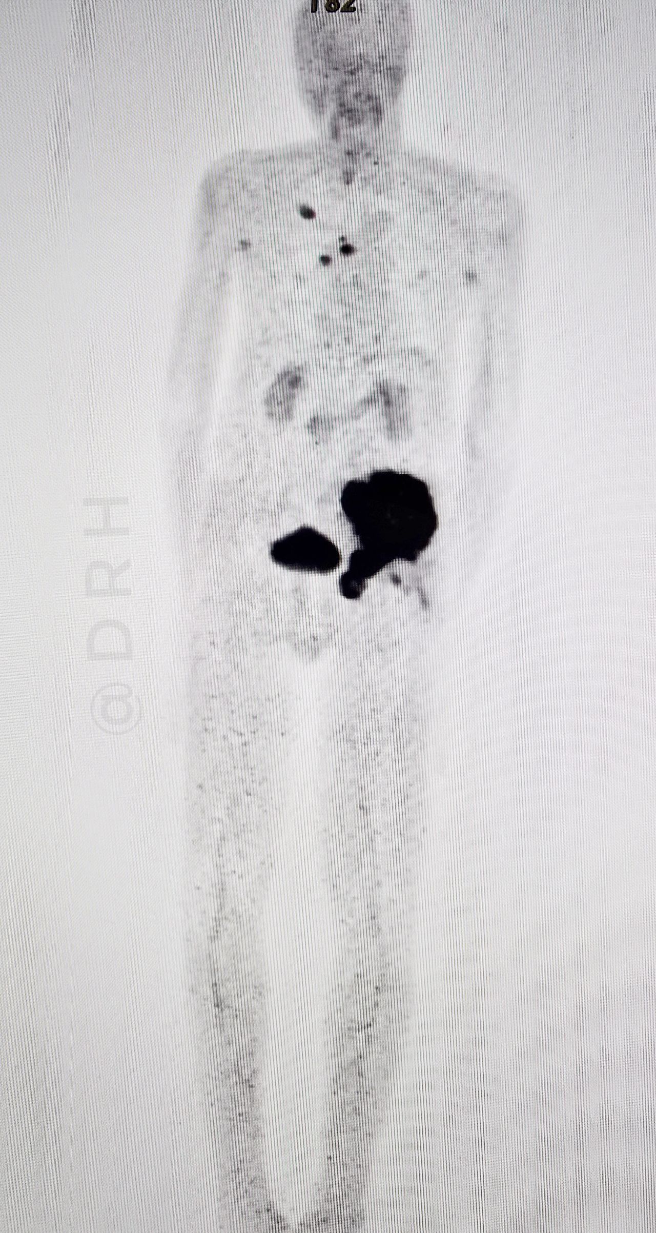
“We recently encountered a case of left iliac bone lytic lesion detected on CT scan highly suspicuous for malignancy who was sent for PET-CT to identify the primary tumor.
A FAPI PET-CT scan was performed, which revealed a primary lung lesion in the right upper lobe along with mediastinal lymph node metastases, confirming the diagnosis of metastatic lung cancer.
This case highlights the value of FAPI PET-CT in oncology in:
– Detecting occult primary tumors.
– Improving diagnostic confidence and staging.
– One of the unique advantages of FAPI PET-CT is its lack of physiological uptake in the brain, which allows for clear detection of brain metastases.Consent- Taken before posting the images.”

Read OncoDaily’s special “Metastatic Lung Cancer: Causes, Symptoms, Treatment, and 2025 Advances in Therapy”.
