Harish Nagaraj, In Charge of the Integrated Molecular Imaging Centre at the Kenyatta University Teaching, Referral, and Research Hospital, shared a post on LinkedIn:
“Case Highlight: Metastatic Carcinoma of Unknown Primary [CUP]
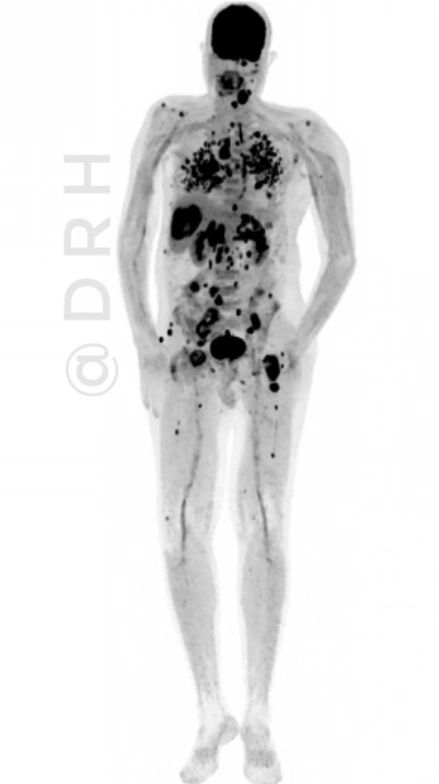
We recently evaluated a complex case of metastatic carcinoma of unknown origin, referred for ‘FDG PET/CT’ to help localize the likely primary site and assess the full extent of disease.
Key PET/CT Findings from the scan:
- FDG avid left hilar mass– suspicious for primary lung malignancy.
- Extensive nodal involvement, including:
- Lower cervical
- Mediastinal
- Retroperitoneal (porto-caval, peripancreatic, hepatic hilar, retrocrural, mesenteric)
3. Visceral and skeletal metastases:
- Large necrotic FDG-avid nodule in the right lobe of liver.
- Innumerable FDG avid nodules in both lungs.
- Bilateral FDG avid adrenal masses.
- Skeletal and soft tissue involvement including intramuscular deposits.
The FDG PET/CT strongly suggested a ‘left hilar primary lesion’ with widespread nodal, visceral, skeletal, and soft tissue metastases.
This imaging modality plays a crucial role in narrowing down the likely origin and guiding further precision diagnostic steps such as tissue biopsy and molecular profiling.
This case underscores the value of FDG PET/CT in the evaluation of ‘carcinoma of unknown primary (CUP)’— not only to identify potential primary sites but also to map the full metastatic burden, essential for treatment planning.
Consent: Obtained before posting.”

More posts featuring Harish Nagaraj.
